DEXIS OP 3D LX
By DEXIS
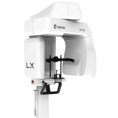
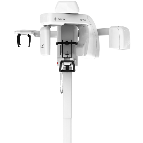
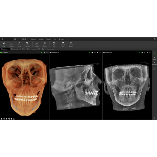
The DEXIS OP 3D LX is the next generation of DEXIS cone beam technology. This 2D/3D imaging platform expands your diagnostic capabilities with a wide range of clinical applications that support your evolving practice and enhance diagnostic confidence.
This next generation system offers flexible field of view (FOV) options ranging from 5x5 cm up to 15x20 cm (optional) – which is the largest view option available on a DEXIS OP 3D platform to date. With shorter scan times, the OP 3D LX captures the maxillofacial complex and large diagnostic areas in one non-stitched scan for fast workflows.

FLEXIBLE FIELD OF VIEW (FOV) SIZES

FULLY UPGRADEABLE

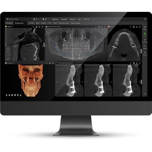
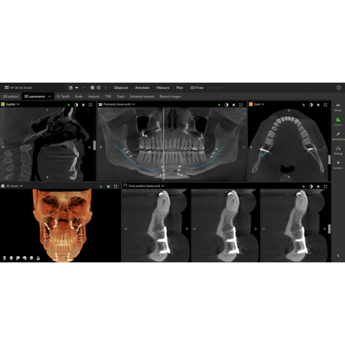
HIGH-QUALITY IMAGES
